
Heart Imaging AI May Change Scans Forever, No Intrusive Contrast Jabs Needed
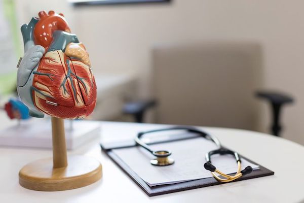
Image via ID 209659879 © Kanpisut Chaichalor | Dreamstime.com
Artificial intelligence (AI) can be used for virtually anything, it seems, and this new discovery by the University of Virginia may lead to breakthrough healthcare.
Although it’s only been tested in one common heart condition, its success signifies the potential to cover many more. And after that, developments to similar technology may see more parts of our anatomy benefitting from using AI in medicine.
This new technology, called virtual native enhancement (VNE), allows doctors to scan patients’ hearts for scar tissue without needing to use contrast injections, which are usually required for effective MRI imaging.
These can be used to monitor hypertrophic cardiomyopathy, the most prevalent genetic heart condition, according to the University’s news release.
The scans are shown to produce images of higher quality than the conventional MRI, and also allow doctors to image the heart more frequently. Plus, it will allow patients who are unable to take contrast injections to finally undergo a heart scan.
Two layers are utilized in this technology. T1 maps of heart tissue, produced by MRI, are enhanced using AI. The maps are combined with MRI “cines,” which are described as “movies of moving tissue”—the beating heart. Together, these result in the VNE image.
Published in the journal Circulation, the team details that the “deep learning model for generating VNE” utilizes “multiple streams of convolutional neural networks” to improve existing T1 maps, which are used to study myocardial tissue.
While tested on patients with hypertrophic cardiomyopathy, the imaging technology has great potential to be used for other heart conditions.
“This is a potentially important advance, especially if it can be expanded to other patient groups,” said researcher Dr. Christopher Kramer of the University. “Being able to identify scar in the heart, an important contributor to progression to heart failure and sudden cardiac death, without contrast, would be highly significant.”
It has “enormous potential” to significantly improve clinical practice, reduce scan time and costs, and expand the reach of [cardiovascular magnetic resonance] in the near future,” the team writes in their paper.
[via University of Virginia, image via ID 209659879 © Kanpisut Chaichalor | Dreamstime.com]
Also check out these recent news


