
Wearable Ultrasound Patch Provides Patients With ECGs On The Go
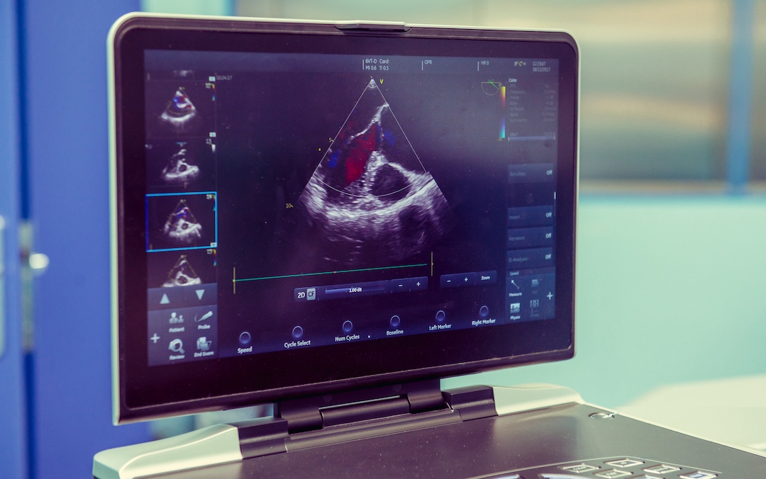
Having a healthy heart is quite literally a vital necessity when it comes to living a full life. However, for those suffering from heart problems, it can be a tricky organ to monitor because patients would need to be hooked up to multiple wires for an electrocardiogram (ECG) or sent for ultrasounds to capture data.
Researchers at the University of California San Diego are developing a new way of examining organs with a wearable ultrasound that acts as a window into the heart. The findings were presented in the nature journal.
The device comprises pressure-powered piezoelectric transducers that can produce deep-tissue imaging. The patch is about the size of a postage stamp and converts electric signals into ultrasound waves to create an image.
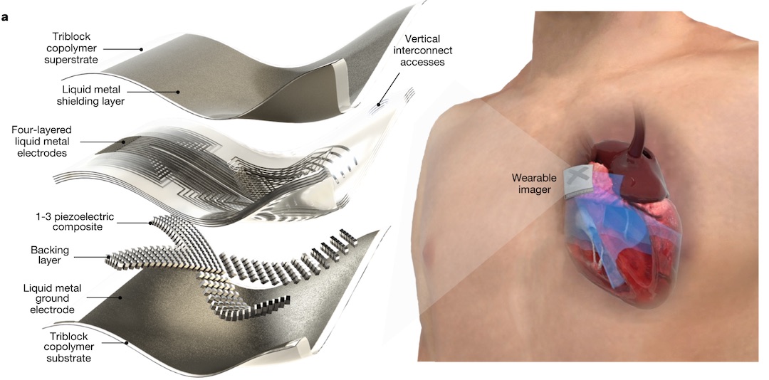
Previous ventures into wearable ultrasounds often used a thin metal film to capture the image. This time the team has employed a stretchable liquid metal electrode to ensure the vision can stay close to the skin. MIT has also created its own version as a sticker that can be placed on the body.
What’s great about this device is that users can wear it while moving about. In addition, it calculates ventricle volume and can provide performance stats. Aside from this, the team hopes to produce continuous metrics for patients with heart disease or those in critical condition.
On top of this, the concept is not limited to just the heart. The team believes it can also work for the spine, liver, and kidneys.
For now, the prototype must be linked to a computer for an image to be captured. However, the results during a test run of the device looked promising as the patch delivered ultrasound pictures close to what would be caught in the clinic.
[via Engadget and New Scientist, images via various sources]


